

Sponsored by:

Innovative Al Techniques for Accurate Analysis of Cell Morphology and Viability
Accurate segmentation of cell boundaries is the first and most critical step underlying the quantification of cell morphology.
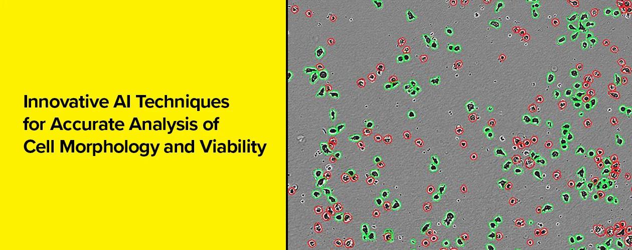
Accurate segmentation of cell boundaries is the first and most critical step underlying the quantification of cell morphology. Label-free imaging enables cell behavior to be monitored in a non-perturbing environment, yet images can be challenging to analyze. Cells in culture have widely varying and fluid morphologies with certain types of cells appearing almost transparent; they grow from sparsely populated to highly confluent. In such a dynamic system, continuously and accurately detecting the boundaries between each cell presents a challenge for traditional computer vision approaches. Here we demonstrate the advantages of using a widely trained neural network for segmentation of different cellular models and using the resulting morphological descriptors to classify cells using a machine learning-based classifier.
The use of label-free imaging for long-term cell monitoring is a powerful approach for examining live cells, providing longitudinal insight into cell behavior, function, and response to external treatment or stimulus.
Key Benefits:
- Improved segmentation enables more accurate morphology classification.
- Microglia viability and activation can be continuously monitored with label-free analysis.
- Non-toxic drug effects can be quantified.
- Transient morphology indicates ferroptotic cell death.