

Sponsored by:

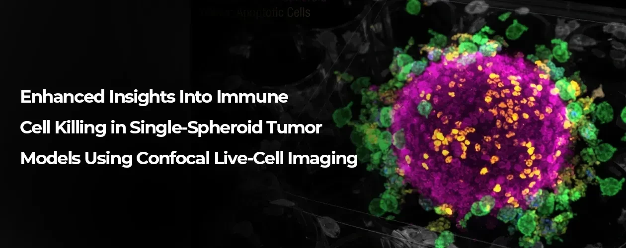
Enhanced Insights Into Immune Cell Killing in Single-Spheroid Tumor Models Using Confocal Live-Cell Imaging
Advanced Confocal Live-Cell Imaging for Immuno-Oncology Traditional endpoint assays miss the dynamic, spatial interactions crucial for evaluating solid tumor immunotherapies.
Advanced Confocal Live-Cell Imaging for Immuno-Oncology Traditional endpoint assays miss the dynamic, spatial interactions crucial for evaluating solid tumor immunotherapies. The Sartorius Incucyte® CX3 Live-Cell Analysis System bridges the gap between in vitro screening and in vivo models. It provides continuous, multi-plane spinning-disk confocal imaging inside your incubator, tracking tumor destruction and immune cell kinetics without disturbing your cultures.
Overcome 3D Tumor Microenvironment Challenges
Solid tumors have complex architectures and gradients that widefield methods blur or merge. The Incucyte® CX3 resolves these limitations with deep-tissue optical sectioning directly where your cells grow.
Deep Optical Sectioning: Achieve a penetration depth of 50–100 µm (~5 cell layers) to resolve dense spheroids.
True Multiplexing: Single-bandpass emission filters eliminate spectral crossover for reliable multi-channel tracking.
Non-Perturbing Reagents: Mix-and-read, no-wash immunocytochemistry labeling preserves cell viability over days.
Versatile Applications in Immuno-Oncology
This flexible workflow delivers rich kinetic data across multiple therapeutic paradigms:
T-Cell Engagement: Differentiate killing profiles between non-activated and pre-activated PBMCs across various E:T ratios.
CAR-T Potency Profiling: Characterize how tumor morphology impacts engineered T-cell infiltration and destruction.
ADCC & ADCs: Quantify NK cell-mediated cytotoxicity triggered by monoclonal antibodies and complex antibody-drug conjugates.
Flexible Screening: Run 3D assays in 96-well or automated high-throughput 384-well formats.
Automated Metrics: Software segments and tracks target cell area loss alongside effector cell proliferation.
Downstream Integration: Couple the non-perturbing assay with HTS flow cytometry for deep phenotypic profiling.