

Advanced 3D Cell Models: Moving from Promise to Practice in Drug Discovery
Nicola Bevan, Manager of Cell Imaging Applications, BioAnalytics Product Development, Sartorius
Advanced 3D cell models have been hailed as the future of drug discovery for more than a decade, yet their routine use in pharmaceutical research remains surprisingly limited. As organoids, spheroids and complex co-culture systems become increasingly sophisticated, the challenge is no longer proving their biological relevance but demonstrating their practical value. To achieve mainstream adoption, the industry must address reproducibility, scalability and data interpretation while ensuring these models deliver actionable insights that improve R&D decision-making.
Introduction:
Over the past decade, organoids, spheroids and other advanced 3D cell models have transformed how researchers approach disease modelling and drug development. Their ability to better reflect human biology than traditional 2D cultures has generated significant enthusiasm across pharmaceutical research, yet widespread adoption remains a work in progress.
The Next Milestone: Mainstream Adoption
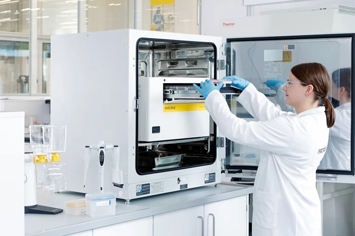
If advanced 3D cell models are to become an established part of mainstream pharmaceutical research within the next five years, three critical areas must mature simultaneously: access to high-quality biological materials, reproducible workflows, and the ability to generate robust quantitative data at scale. Researchers need reliable access to relevant cells, tissues and matrices, supported by clear sourcing, consent and intellectual property frameworks. Equally important is a more sophisticated understanding of reproducibility and analytical capability through automated imaging, multiparametric analysis and AI-enabled data interpretation. In practice, this will require a shift from treating each model as a bespoke research project toward developing dependable platforms that can be transferred between laboratories. Clear standard operating procedures, defined passage limits, validated readouts and appropriate positive and negative controls will help reduce uncertainty. It will also be important to understand which elements of a model must remain fixed and which can be adapted to suit a particular disease area or screening objective. This balance between standardisation and biological flexibility will determine whether advanced models can be used consistently across discovery, translational research and preclinical development.
Where Expectations Still Exceed Reality
Despite the excitement surrounding organoids and other advanced models, several common assumptions remain ahead of current capabilities. Throughput remains challenging, access to patient-derived organoids is uneven, and researchers still need stronger links between model phenotypes, functional biology and clinical outcomes. Another limitation is that complexity does not automatically translate into better prediction. A model may contain several cell types and reproduce aspects of tissue architecture, yet still lack the immune, vascular or metabolic interactions needed to answer a particular question. Researchers must therefore define the intended use before selecting the model format. In some cases, a simpler spheroid with a well-characterised endpoint may be more informative than a highly complex system that is difficult to control. The most useful approach is to match model complexity to the biological decision being made.
Learning from Early Successes
Commercial tissue models used in skin and ocular research have demonstrated considerable value in toxicity, irritation and barrier-function studies. Oncology has also seen encouraging progress through tumour spheroids and tumour-immune co-culture systems. Across all successful applications, deep phenotyping, relevance and characterisation have proven essential. These examples also demonstrate the importance of selecting endpoints that are directly linked to the intended application. In toxicity studies, changes in barrier integrity, tissue structure or inflammatory signalling may provide more useful information than viability alone. In oncology, drug response can be evaluated through changes in spheroid growth, invasion, cellular composition and interactions between tumour and immune cells. Combining several complementary endpoints can provide a more complete picture of treatment response and reduce the risk of drawing conclusions from a single measurement.
Addressing the Challenges Often Overlooked
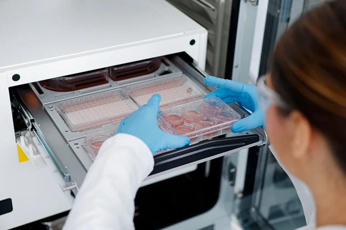
Operational challenges including the transport of living tissues, development of animal-free automation-compatible matrices, and analysis of thick tissues deserve greater attention. Standardised logistics, advanced matrix technologies and improved imaging methods will be critical for scaling adoption. Sample handling is another practical consideration. Variations in temperature, transit time, mechanical stress and recovery conditions can influence tissue health before an experiment even begins. These effects may be especially significant when working with fragile patient-derived materials. Establishing defined procedures for collection, shipment, receipt and equilibration will therefore be as important as optimising the assay itself. In parallel, laboratories will need instruments and consumables that support consistent processing without compromising tissue structure or cell viability.
Rethinking Variability
Variability should be viewed as biological information rather than noise. Patient-derived models offer valuable insights into human diversity, making robust controls, multiparametric analysis and longitudinal measurements increasingly important. The challenge is to distinguish meaningful biological variation from technical variation. This requires careful experimental design, sufficient biological replicates and the use of reference samples where appropriate. Recording donor characteristics, culture history and assay conditions can help researchers interpret differences more accurately. Rather than reporting only average responses, advanced analysis should also identify responder and non-responder populations, subgroups with distinct phenotypes and changes that occur over time. Such information could be particularly valuable for understanding patient stratification and the potential mechanisms of treatment resistance.
The Growing Role of AI
Artificial intelligence will be indispensable for analysing the complexity of advanced 3D models. Applications range from image analysis and phenotypic classification to linking experimental datasets with donor and clinical information, while supporting future automation of culture workflows. The value of AI will depend on the quality and diversity of the data used to train and evaluate these systems. Models developed from a narrow set of donors, laboratories or imaging conditions may perform well in one setting but fail when applied elsewhere. Independent validation, careful annotation and ongoing monitoring will therefore be necessary. Researchers will also need to maintain appropriate human oversight, particularly when AI-generated classifications influence decisions about compound progression or safety. Used responsibly, AI can reduce analysis time, identify patterns that are difficult to detect manually and make high-content experiments more manageable.
Looking Ahead
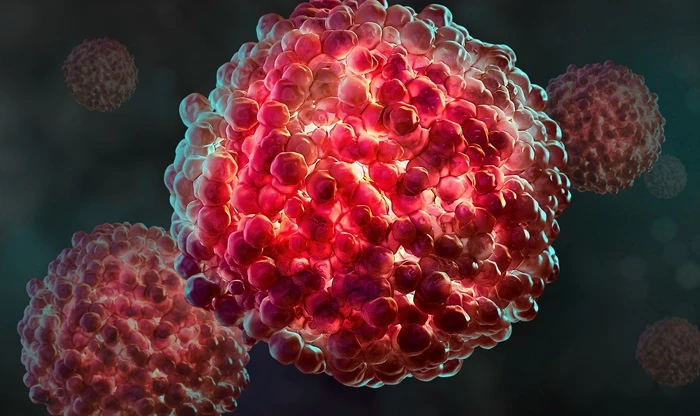
Over the next five years, tissue toxicity and oncology are likely to benefit most from advanced 3D models. Long-term success will depend not on complexity alone, but on delivering standardised, reproducible and biologically relevant models that support meaningful R&D decisions.
From Model Development to Decision-Ready Evidence
A major step toward practical adoption will be the development of decision frameworks that define what a 3D model is expected to contribute at each stage of the drug discovery process. A model used for early compound ranking may not need to reproduce every feature of a human tissue, but it must provide a consistent and sensitive readout that distinguishes meaningful biological activity from experimental variation. Conversely, a model intended to support candidate selection or safety assessment will require stronger evidence of translational relevance, including comparisons with reference compounds, primary human data and, where available, clinical observations.
This makes model qualification just as important as model construction. Researchers will need to establish clear acceptance criteria for cell identity, tissue architecture, viability, functional maturity and assay performance. These criteria should be assessed over time rather than at a single endpoint, because many 3D systems evolve during culture. Longitudinal monitoring can reveal whether a model is becoming more physiologically relevant or drifting into an unstable state. It can also help identify the optimal window for treatment and measurement, improving both data quality and comparability between experiments.
Making Complex Biology Easier to Measure
Imaging will be central to this transition. Conventional endpoint assays often require the destruction of a sample, limiting the amount of information that can be collected from each model. By contrast, live-cell imaging can follow growth, morphology, movement, cell death and treatment response in the same sample over several days. When combined with fluorescent reporters and multiparametric analysis, these measurements can reveal subtle changes that would otherwise be missed. For example, a compound may not immediately reduce spheroid size but may alter cell organisation, induce selective cell death or prevent invasive outgrowth. Such effects can be important indicators of mechanism and therapeutic potential.
However, more data will only be valuable if it can be interpreted consistently. Image-analysis pipelines therefore need to be transparent, robust and adaptable to differences in sample size, matrix composition and imaging conditions. Automated segmentation and feature extraction can reduce user bias, while appropriate controls and quality checks help ensure that algorithms are not simply detecting changes in illumination, focus or sample position. The goal should not be to replace scientific judgement, but to make complex observations easier to quantify, compare and communicate.
Building Confidence Across Organisations
Collaboration will also be essential. No single laboratory is likely to solve every challenge associated with advanced 3D models. Partnerships among pharmaceutical companies, academic groups, technology developers and contract research organisations can accelerate the creation of shared protocols, reference materials and benchmarking datasets. Common reporting standards would make it easier to compare results generated using different cell sources, matrices, instruments and analysis methods. They would also support regulatory discussions by providing a clearer description of how a model was developed, qualified and applied.
Ultimately, the strongest 3D models will be those that fit naturally into existing research workflows. They must be practical to culture, compatible with automation, supported by reliable analytical tools and capable of producing results within the timelines required for drug development. Complexity should be introduced when it answers a specific biological question, not simply because a more elaborate model is technically possible. By focusing on measurable value, advanced 3D systems can move beyond their reputation as promising research tools and become dependable platforms for better, faster and more informed pharmaceutical decisions.

Nicola Bevan is a Manager of Application Development within the BioAnalytics Application Development team at Sartorius. She is actively involved in the research and development of novel reagents and applications, enhancing the capabilities of life science instruments such as the Incucyte® live-cell analysis system and the iQue® HTS cytometer. With over 30 years of experience, Nicola is a highly skilled cellular pharmacologist. She has an extensive background in the Pharmaceutical and Biotech industries, where she has gained broad experience in large pharma industrial screening departments and the support of early drug delivery programs.